What Can I See On A 8 Week Ultrasound? Things To Expect!
Every stage of your pregnancy brings a new challenge in front of you, and along with a glow in your face comes side effects. Around 8 weeks, your baby grows from a tiny seed to a humanoid form.
By the end of week 8 of your pregnancy, your baby has grown about an inch and weighed about half an ounce. But his internal organs, like the heart, lungs, and central nervous system, are nearly full grown.
The baby’s heart will be beating at a good rate of 160 beats per minute by now, and it already has four chambers. That is the heart rate that an adult has after one hell of a workout.
What Is An Ultrasound?

An ultrasound or diagnostic sonography is a type of digital imaging method that utilizes sound waves to create images of the structure inside the body. Through these images diagnosing the internal organs is done easily.
When you are in an 8-week pregnancy, it is recommended that you take at least one 8-week ultrasound. It is very important to have a prenatal ultrasound, as it shows the fetus’s position and shape inside the uterus. And ultrasounds are also painless.
Through an ultrasound, your doctor can check whether the fetus has any problems or difficulties. Before labor, these assessments are very important.
Based on what your doctor wants to look for, there are two types of ultrasound: transabdominal and transvaginal.
Through a transabdominal ultrasound, your doctor basically takes an image of your fetus. First, your doctor will apply gel to a wand-like instrument called s transducer. Then the device will be moved across your belly to take images.
With a transvaginal ultrasound, your doctor does the same as before, but this time the transducer goes inside your vagina to take the images.
Purpose And Importance Of An Ultrasound:

The main purpose of an ultrasound is to determine and check on the fetus’s position and shape. However if you are going to take early 8-week ultrasound pictures, then it will be too early to define the legs and arms of the fetus. Even too early to determine the sex of the baby.
An ultrasound, as early as 8 weeks, helps to determine other important factors, such as the fetal heart rate. Other factors, such as fetal development and the fetus’s gestational age, are also determined. An 8-week twin ultrasound may also be used to determine if you have twins.
During your 8-week pregnancy, the ultrasound can help you determine certain pregnancy-related factors such as the location of the fetus, age, heartbeat, and also if you have more than one baby. If you are experiencing some unusual symptoms, then the doctor can even check for ectopic pregnancy as well.
If you want to have an 8-week ultrasound 3D, it will give a more detailed and 3-dimensional image of your baby. However, it is recommended more after the second or third trimester, as it provides a memorable memento for the parents.
The 6 Week Ultrasound

You should always visit your doctor for a 6-week ultrasound appointment if you have certain health risks. Here are a few reasons why doctors recommend a 6-week ultrasound to expect mothers.
- Age (if the mother is over 35, pregnancies are considered high-risk)
- History of pregnancy complications (such as miscarriage or blood loss during pregnancy)
- Medical history (such as diabetes, high blood pressure)
The 8 Week Ultrasound:

During week 8, the ultrasound you recommended is a transabdominal one instead of a transvaginal ultrasound.
During an 8 week ultrasound, there is not much to determine other than the heartbeat, position, and size of the fetus. In the eighth week, the fetus is more developed; the yolk sacs, fetal pole, the embryo, and heartbeat are defined.
If you don’t know what a yolk sac is, it is a protective sac that holds the amniotic fluid. It provides nutrients for the fetus.
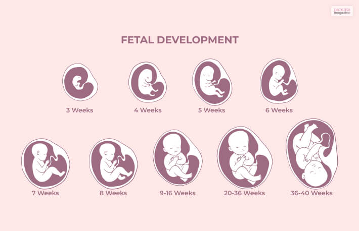
At this stage, don’t expect your baby to look like a human. It will be in the shape and size of a kidney bean. You may see forms of arms and legs, but nothing prominent. It is also possible that you can see more than one embryo or multiple.
Your doctor will recommend you to take a 6 to 8 week ultrasound, even though it’s your first trimenter, to check for signs of a healthy pregnancy.
Difference Between Transvaginal & Abdominal Ultrasound
Transvaginal ultrasound is quite different than abdominal ultrasound. An abdominal ultrasound involves a technician using a wand to take an ultrasound of the stomach. It usually takes thirty minutes or so.
But a transvaginal ultrasound is a little different. In this case, the wand is inserted inside the vagina to check the growth and the development of the fetus. This is done during the early pregnancy.
This ultrasound goes beyond checking the heartbeat. The technician can check the baby’s different features. For example, the gestational sac and the fetus’ crown-rump lenghth. These features help us understand the gestational age of the fetus. Also, they can help pinpoint the due date.
Fetal Development

Towards the end of week 8, your uterus has grown to the size of a lemon, and not a small one. Your little fetus is also growing by getting all the nourishment from the yolk sac inside the amniotic sac.
Slowly the placenta will develop, which sticks to the uterine wall and provides nutrients to the growing baby. At this stage, your fetus is about half an inch in size. The heart is already developing, and you can even see the forms of arms and legs.
Other organs like the eyes, ears, liver, and lips are all starting to form and take shape. At this stage, you are able to hear a strong, healthy heartbeat.
The umbilical cord is already formed that connects the placenta with the fetus. Even though the fetus is only taking a human shape, the head is bigger than the body, so don’t find this weird during your 8 week ultrasound.
Frequently Asked Questions! (FAQs):
Being an expecting mother, you are definitely bound to have more questions; here is what other mothers like you asked.
Ans: It is highly possible that some abnormalities in the fetus or the embryo can be detected as early as 7 to 8 weeks.
Ans: 8 weeks is considered a milestone as this is when you can see and hear the heartbeat of the fetus. This is the main reason why the doctors wait until week 8 for an ultrasound.
Ans: An 8-week old developing fetus would look like a kidney bean and only half an inch taller. The fingers and limbs are definitely growing and are visible. Eyes, ears, and other organs are also forming.
Wrapping Up!
It is very important to get an ultrasound during the early stages of pregnancy, as it helps the doctor and you as parents to check up on the fetus. An 8 week ultrasound is able to see the process of the growing fetus and look for signs of any abnormalities.
For any expectant mother, it is important to have monthly checkups from the doctor and take extra care of themselves.
Hope this article is proven helpful for you, and if you have any more questions, feel free to ask, I will surely get back to you.
Read Also:
Leave a Reply
All Comments
Already have an account?
Sign In
Create your account
User added successfully. Log in


7th June, 202425 July 2023 at 10:08 AM |
Fascinating blog!